
How do we See? The Stunning Structure of Our Eyes
People used to believe for ages that our eyes emit rays of light, and that is how we see things around us. But that was until an Arab scholar named Ibn al-Haytham corrected that mistake by arguing that light is reflected from objects around us and then bounces off into our eyes, and that’s indeed how we see things.
Our eyes are a very complex and invaluable structure that we all need to know more about. So in this article, we are going to dive deep into our eyes, see how they work, what they are composed of, and finally, some visual defects that might happen in our eyes.
But before we get into how we see things and how vision works, we need to know first the components of our eyes.
Table of content:
- The components of the eye.
- How do we See Things?
- Why do we Need a Brain to See?
- Visual Defects.
- Short Quiz on the topic.
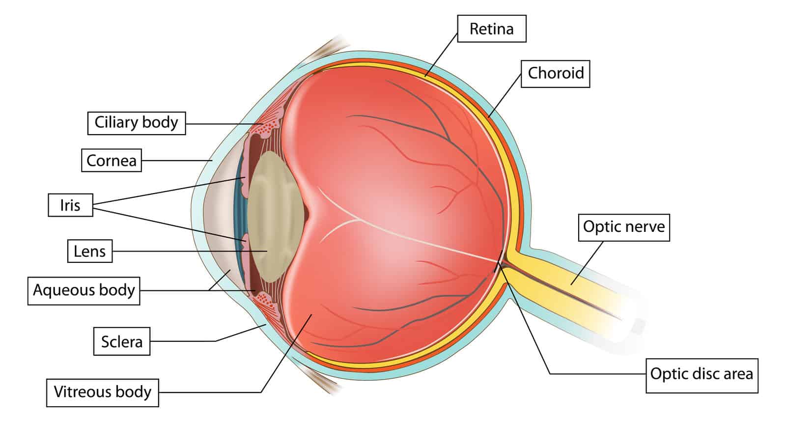
The Components of The Eye
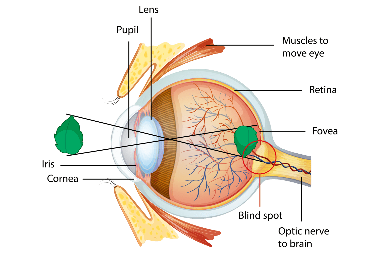
If we take a vertical cross-section of the human eye, we will find the structures listed below from the front backwards.
The Cornea
It is the clear, outermost layer of your eye. It looks exactly like a transparent contact lens. It helps you focus the light so that you can see clearly. The cornea of your eye is so unique in different ways. For example, unlike all other organs in your body, it does not have a blood supply. Instead, It receives nutrition and oxygen directly from the air and the fluid that fills the eye. That adds to the clarity of the image you see; if we were to have blood vessels crossing our corneas, we wouldn’t be able to see clearly.
The Sclera
This is the white outer layer of your eye. It is the backward continuation of your cornea. The cornea and the sclera together form the outer protective layer of our eyes. The sclera is very important as it may turn yellow if you get jaundice (a problem in your liver).
The Iris
This is the part that gives your eyes their colour. It has two sets of muscles that control the diameter of the hole in the middle of the iris (the pupil).
These muscles are:
The Dilator Pupillae Muscle
As its name implies, it causes dilatation (widening) of the pupil opening to let more light in. This happens in dark places where you can barely see things to help you navigate your way without getting stuck.
The Constrictor Pupillae Muscle
As you may have guessed, this muscle does the opposite, as its name also implies. When it contracts, it limits the amount of light passing through the pupil opening by narrowing it. That is what happens when you spend some time out on a sunny day or in a very bright place.
Your pupils constrict so as to protect your eyes from excess light that might be harmful to your eye. You might have noticed that when you spend some time in the sun and then move to a darker place, you struggle to see what is inside that place for a few moments, and then you start to see things clearly.
Well, that is because when you are in a very bright place, your pupils constrict and limit the amount of light that goes into your eyes. And then, when you move to a darker place, your pupils take a few seconds to adapt and change their diameter to allow more light in to be able to see things clearly in that dark place.
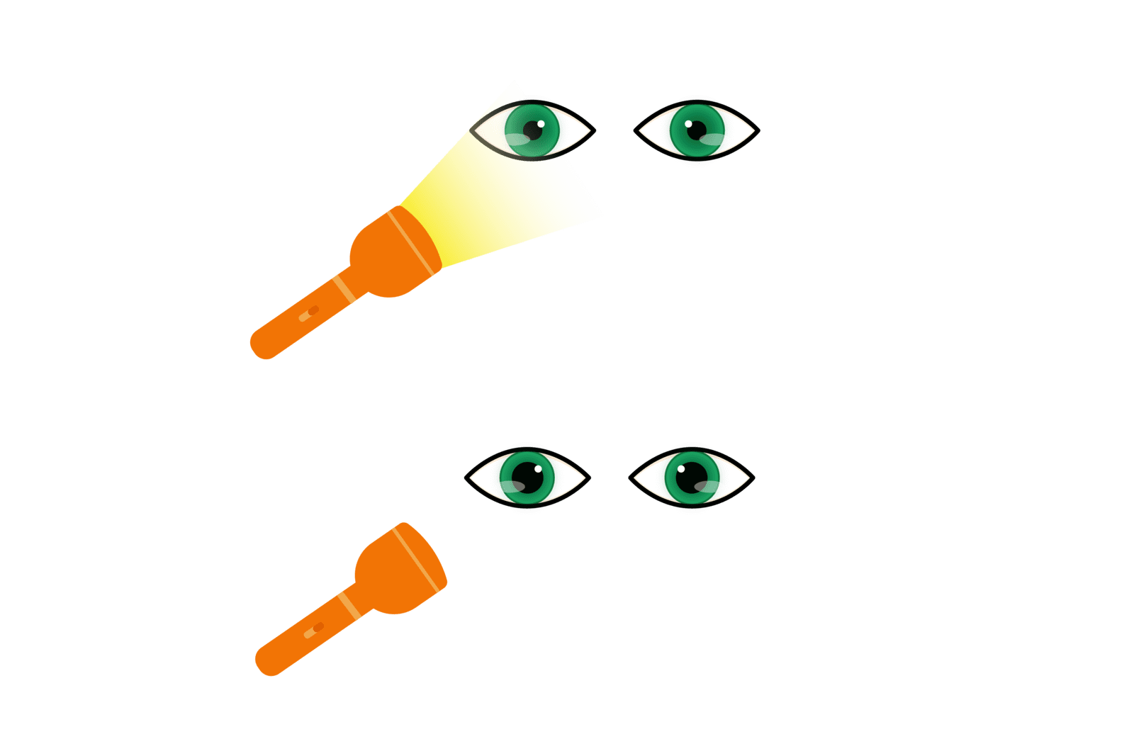
Want to Test Your Pupillary Muscles?
Let’s do that experiment together.
- First, bring a torch or use your mobile phone torch.
- Then, stand in front of a mirror in a well-lit room (not too bright) and look into your eyes.
- Try to observe that hole in your iris.
- Focus the light of the torch on your eyes.
- Observe how that hole becomes smaller and smaller as you bring the torch light closer to your eye. And how it gets bigger (dilates) as you move the light away from your eyes.
- And here you go, you have just done a very famous test called “The light reflex”. Your doctor might do that test to make sure your eye and its nerve supply are okay.
That is the light reflex:
Notice when the torch is pointed at your eyes, your pupils constrict. When it is turned off or moved away, your pupils dilate.
The Pupil
This is the hole that you can see in the middle of your iris. Its diameter is under the control of the muscles mentioned above.
The Lens
It is a clear lentil-shaped structure inside your eye that works with your cornea to focus light rays coming out of an object on the retina.
The Ciliary Body
This part of your eye contains a tiny muscle called the ciliary muscle that connects to your lens by some thin filaments called the suspensory ligaments of the lens. These filaments help hold the lens in its place. The ciliary muscle controls the curvature of your lens and, therefore, its power.
When the ciliary muscle contracts, the suspensory ligaments become loose. That, in turn, causes your lens to get more convex (increase the curvature of your lens), which means increasing the power of your lens and vice versa.
You need to increase the curvature of your lens (its power) when you are reading a book or focusing on something close to you. But to be able to see things far away from you, you need to relax your ciliary muscle so as to decrease the lens curvature (power).
The Choroid
This layer provides blood supply to your eye’s inner and outer layers. That is why it is located in the middle between the outer layer (formed by the cornea and sclera) and the inner layer (formed by the retina).
The Vitreous Body
This transparent jelly-like structure keeps your eyes rounded and prevents them from collapsing.
The Retina
This is the innermost layer that lies at the back of your eye. It is also a light-sensitive structure that contains special cells called photoreceptors. These photoreceptors can turn the light they receive into electrical signals.
There are two main types of cells inside your retina: the cones and the rods. Cones are responsible for colour vision. Rods are involved in detecting movements, and they only give the brain white and black images.
The Optic Nerve
This is a cord-like structure that transmits electrical signals from your retina to your brain, especially to the occipital cortex (an area that lies at the back of your brain).
How do we See Things?
Now, after knowing the parts of the human eye, we are very well-equipped to continue the journey and explore how we see things and how our visual system works.
To see an object, the light coming out of that object must pass through specific structures in your eye until it gets transformed into an electrical signal that your brain recognises and translates into a meaningful image. And although that might sound a little bit complex, that process takes only a fraction of a second to happen in real life.
So here is how it happens:
- First, light passes through your transparent cornea with minimal refraction.
- Then, it passes through your pupil.
- After that, it hits your lens, where it gets refracted and focused even more.
- Then, it moves into your eye until it hits your retina.
- Next, the photoreceptors in the retina transform that light energy into an electrical signal that passes to your brain through your optic nerve.
- Finally, in the cerebral cortex (occipital lobe of the brain), that signal gets translated by your brain into a meaningful image.
Why do we Need a Brain to See?
First of all, the image that is formed after light hits your lens is flipped upside down and also reversed from right to left, which means your brain can’t make a meaningful image out of this thing. So, your brain corrects the position of this image to make it meaningful to you. This process takes a fraction of a second, and none of us is even aware of it.
Second of all, your brain not only identifies objects but also gives you more information associated with these objects.
For example, when you see a pen, your brain not only tells you that this is a pen, but it also associates that pen with certain things like writing and paper. That is a very interesting thing to know as in some diseases, where a specific area of the occipital cortex gets damaged, the patient can see things clearly, but they fail to tell what they are or how we use them despite having healthy and perfectly normal eyes.
The problems of the visual cortex are really fascinating to know since the eyes are completely normal and healthy. The problem here is in the visual cortex (the occipital lobe of the brain).
A very interesting defect that occurs as a result of injury to the occipital lobe of the brain is known as visual agnosia. In this weird disorder, patients can see everything around them but can’t recognise what they are looking at. To know more about this strange yet interesting disorder, check out this book by Oliver Sacks, The man who mistook his wife for a hat.
Visual Defects
Have you ever seen someone wearing glasses and wondered why they wear them?
Well, that is because those people have a problem with their eyes. In this section, we are going to discover some problems that might occur in our visual system. And because visual problems are very common and numerous, we will focus only on the most common problems, which are the eye’s refractive errors.
Refractive Errors
Since both the cornea and the lens work together to collect and focus the light rays on your retina, any change in their curvature would lead to a defect in their refractive power. This, in turn, will lead to a refractive error in the eye. As a result, the image your eye receives will no longer fall precisely on your retina, leading to blurry vision that would require eyeglasses to be corrected.
Refractive errors of the eye include two major problems:

Far-sightedness (Hypermetropia).
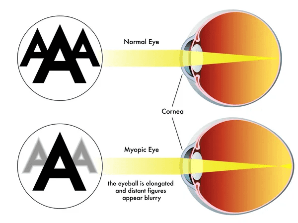
In this disorder, the patient can see far objects clearly but finds it difficult to see near objects. It occurs mainly in people who have short eyeballs with decreased curvature of the cornea or the lens. This makes it difficult for them to focus on near objects.
Near-sightedness (Myopia)
Contrary to hypermetropia, myopic patients can clearly see near objects, but they find it difficult to see far objects. It happens mainly in people with eyeballs longer than normal or whose cornea is so steep (highly curved).
Conclusion
Now, we have discussed the structure of our eyes in detail and talked about the importance of your brain in the process of vision. It’s time to test yourself out. Ready?
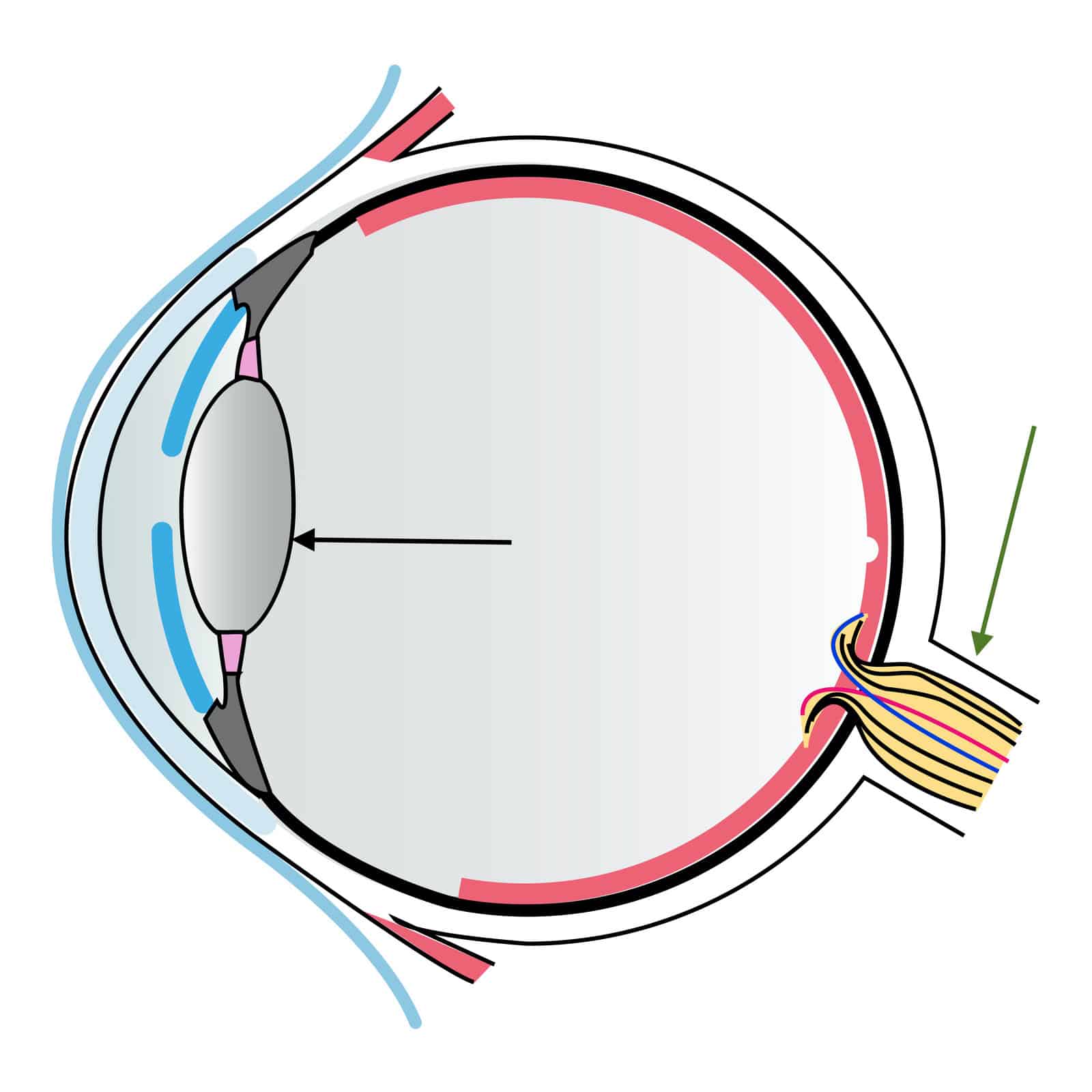
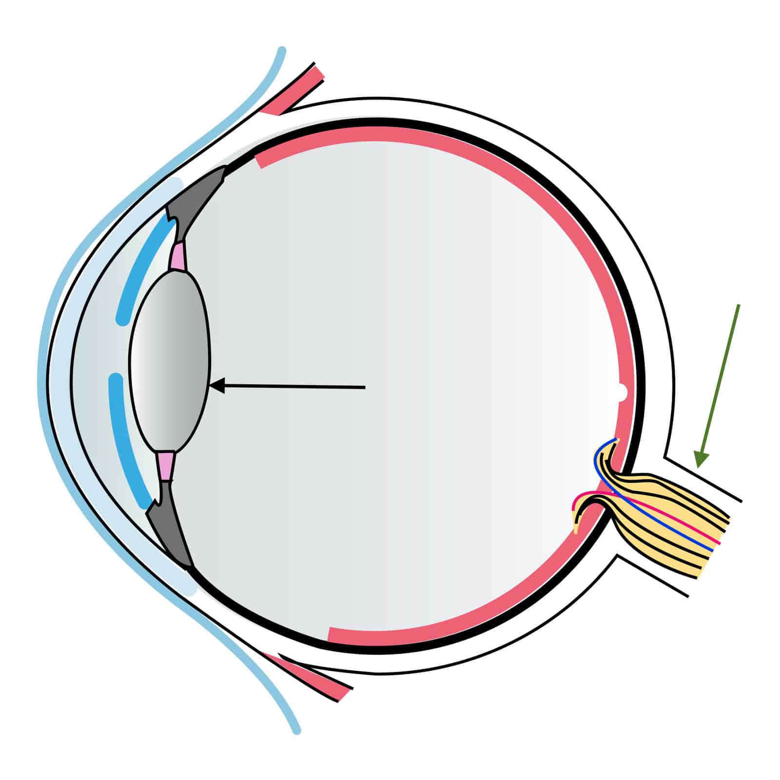
Q1. What is the name of the structure pointed at by the green arrow?
- Retina.
- Cornea.
- Sclera.
- Optic nerve.
Q2. Which structure of these keeps your eye rounded and prevents it from collapsing?
- Lens.
- Cornea.
- Vitreous body.
- Sclera.
Q3. Our eyes emit light rays, and that is how we see things around us.
- Yes.
- No.
Q4. When you are in a very dark place, your pupils dilate.
- Yes.
- No.
Q5. What is the name of the structure pointed at by the black arrow?
- Retina.
- Cornea.
- Lens.
- Optic nerve.
Q6. Which one of these structures of your eye does not have a blood supply:
- Optic nerve.
- Cornea.
- Ciliary body.
- Iris.
Q7. this structure transmits the electrical signals from your retina to your brain:
- Optic nerve.
- Ciliary body.
- Lens.
- Sclera.
Q8. Which of the following pathways best describes the correct order that light travels through your eye:
- The lens – the cornea – the optic nerve – the brain.
- The optic nerve – the cornea – the lens – the brain.
- The cornea – the lens – the optic nerve – the brain.
- The brain – the optic nerve – the lens – the cornea.
Q9. The reason why you need a brain to see is which of the following:
- Make sense of the objects you see.
- Correct the reversed upside-down image formed at your retina.
- Associate the objects you see with meaningful information.
- All of the following.
Q10. Which of the following is incorrect regarding near-sightedness:
- People with this condition can clearly see near objects.
- They find it difficult to see far objects.
- Their eyeballs are longer than the normal average.
- Their eyeballs are smaller than the normal average.
The correct answers:
Q1 – 4
Q2 – 3
Q3 – 2
Q4 – 1
Q5 – 3
Q6 – 2
Q7 – 1
Q8 – 3
Q9 – 4
Q10 – 4
Enjoyed this? Why not check out some fun facts about the nose?
Why not subscribe to our LearningMole Library for as little as £1.99 per month to access over 1000 fun educational videos.


Leave a Reply